

RADMAX







RADMAX
Digital Imaging Software
Smart Diagnostics at Your Fingertips
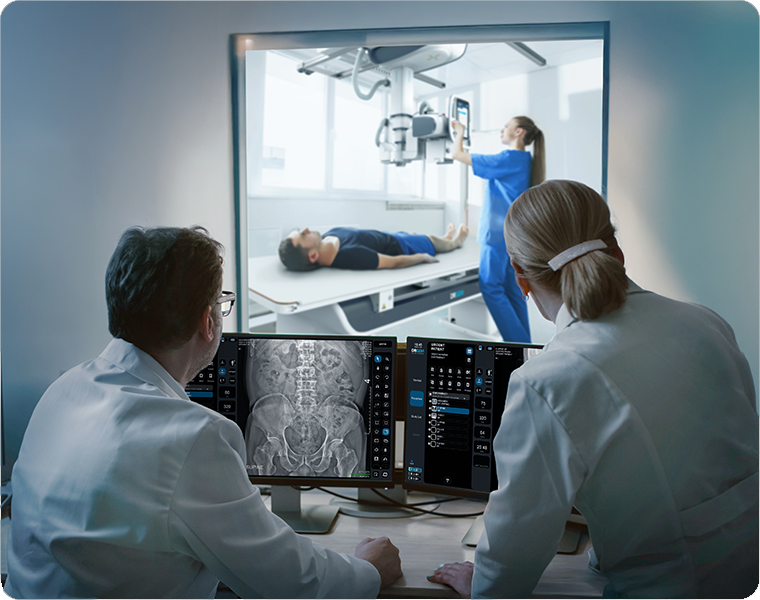
Developed by DRGEM, RADMAX provides top imaging quality at reduced doses, offering retrofit compatibility with a variety of X-ray systems.
With an intuitive interface, RADMAX simplifies workflows by minimizing exam steps and providing high-resolution images with a full suite of diagnostic tools.

Intuitive Touch-Friendly Interface
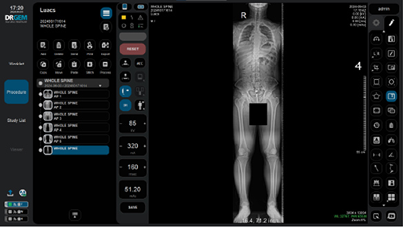
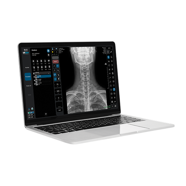
RADMAX offers a seamless touch-enabled UI/UX with optimized screen layouts, maximizing the imaging viewer area and enhancing workflow efficiency for effortless navigation.
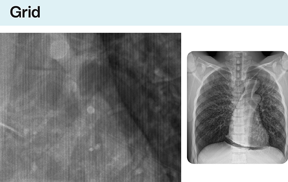
Grid Line Suppression for Enhanced Clarity
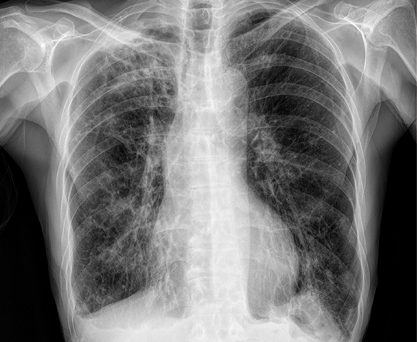
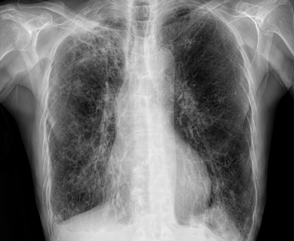
RADMAX minimizes or eliminates grid lines in X-ray images, enhancing overall clarity and sharpness. This advanced feature improves diagnostic accuracy by delivering high-quality, unobstructed imaging for precise evaluations.
Bone Suppression for Enhanced Clarity
The bone suppression feature improves the visibility of soft tissues by reducing the prominence of bones in X-ray images. This enhances image clarity and aids in more accurate diagnoses.
Scatter Reduction for Clearer Imaging
The scatter reduction feature uses a software-generated grid to minimize image noise and enhance contrast, improving diagnostic image clarity without the need for physical grids.
Image Stitching for Comprehensive Visualization
The image stitching feature merges up to five images into a single seamless view, enabling comprehensive visualization and supporting accurate diagnostic evaluations.
AI-Based Diagnostic Assistance
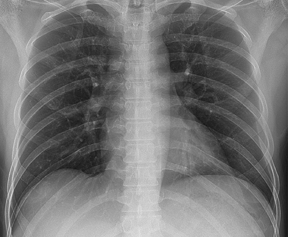
RADMAX integrates AI-based diagnostic assistance to enable precise and immediate detection of suspected abnormalities. This advanced feature enhances diagnostic accuracy and supports faster decision-making in clinical workflows.
Key Features
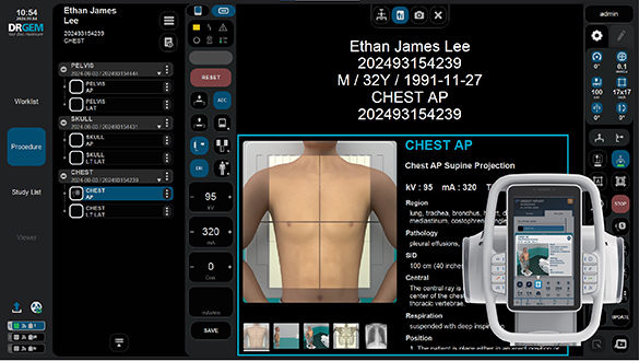
Positioning Guide for Accurate Imaging
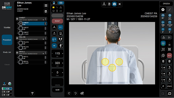
The positioning guide assists operators in aligning patients and the X-ray system with precision, ensuring optimal imaging results. By minimizing retakes and reducing patient radiation exposure, it enhances workflow efficiency and patient safety.
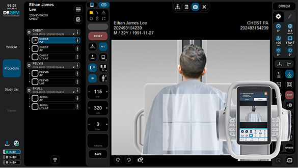
Live Streaming for Enhanced Accuracy
RADMAX supports real-time live streaming via an optional camera, allowing operators to monitor patient status in real time. This feature provides immediate feedback, optimizing the imaging process, improving diagnostic accuracy, and reducing the need for retakes.
Patient Movement Detection
RADMAX includes patient movement detection that alerts operators before X-ray exposure. This feature prevents retakes and ensures the acquisition of high-quality images, enhancing workflow efficiency and diagnostic precision.
Radiation Dose Structured Reports (RDSR)
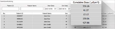
Generates structured reports on patient radiation exposure to support dose management and enhance patient safety.
Virtual AEC Field Alignment
RADMAX enables precise alignment of the AEC (Automatic Exposure Control) field with the patient’s body during positioning. This feature ensures accurate exposure, optimizing image quality and improving diagnostic outcomes.
Exposure Index
Monitors Exposure Index (EI) and Deviation Index (DI) to ensure optimal radiation exposure for high-quality images and enhanced patient safety.
Reject Analysis
Analyzes rejected images to identify patterns and reduce imaging errors, helping to improve workflow efficiency and minimize waste.
Audit Trail
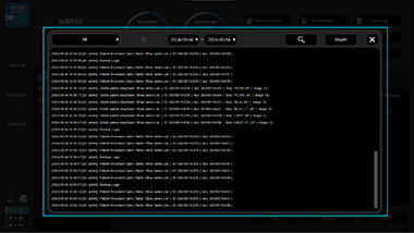
Tracks all system activities to provide detailed logs, ensuring accountability and maintaining compliance with clinical and regulatory standards.
DICOM Transport Layer Security (TLS)
Secures the transfer of sensitive imaging data across networks, ensuring confidentiality and compliance with industry standards.